
X-ray & Ultrasound
Some time ago, we left the traditional black and white gelatin films in the past, where they belong, and are now on our second digital solution the same technology on offer at modern teaching hospitals like the Royal Infirmary of Edinburgh.
As you will know, an x-ray used to be a black and white film and going digital allows many more shades of grey, meaning more can often be seen in the image, compared to a film x-ray.
The first digital systems, called CR or Computerised Radiology used a reusable digital “film” read by a scanning machine line by line and was certainly a step forward. DR or Digital Radiography uses a fixed sensor with millions of tiny sensors to directly capture the image and send it to the computer. To give you an idea a penny would cover over 16,000 sensors each contributing it’s own tiny dot making up a hugely detailed image. All in about four seconds!
What are the Advantages of Direct Digital Radiography?
- Quality of image: Digital Radiography (DR) is currently state of the art and is the gold standard. You see the direct image, whereas a computerised image from the reusable film (CR) is a bit like a photocopy of a photocopy and whilst a step up from films, our digital system produced images, often of jaw dropping quality.
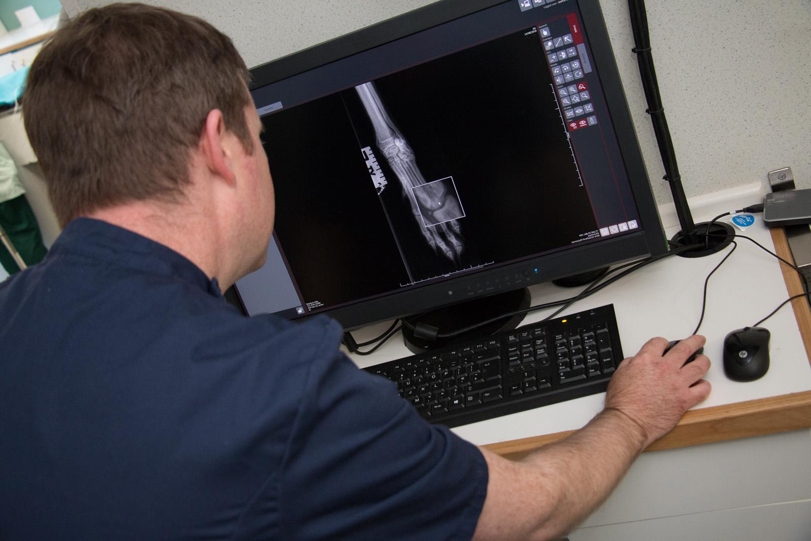
- No delay: As soon as the equipment is turned on, we are ready to take the radiograph. With an old fashioned wet film processor, 15-20 minutes is needed for the fluids to warm up, even longer with manual processing, still seen in some veterinary practices. With our new system, your pet’s image is on the big high-resolution FDA approved viewing monitor.
- Fewer exposures: As the software can view many more greys, a potentially light or dark image is automatically adjusted so we can see what we need to, whereas with a film, what you see is what you get and would need to be repeated. Often a film taken for bone is too dark for the soft tissues and a film taken for soft tissues is too white for bone. With digital you can see both properly, through the contrast controls.
- Lower dose: We have been able to reduce the x-ray settings converting to digital. Although all x-rays involve a minute radiation risk, any reduction has to be welcome.
- Media Sharing Ability: A digital image can be copied to a disc or emailed. This means we can send a copy of your pet’s x-rays to a specialist anywhere in the world within a matter of minutes.
- Environmentally sensitive: We no longer need to buy and store films or buy and dispose of chemicals, which include silver salts which are potentially hazardous to the environment. A saving in things and a saving in fuel for delivery and collection mean a significant reduction in carbon footprint over the long term.
Radiography Examples
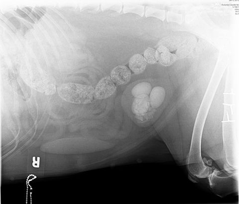
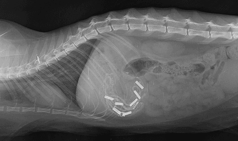
Bladder stones
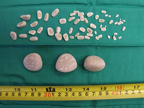
There were more than 60 stones in the bladder of this dog on a CR image. They were removed surgically. The horizontal row of whitish blobs is, in fact, faeces and is normal for a dog fed on raw chicken!

Here are the stones Note the long incisor (front) teeth on the CR image, the big gap and then the molars and how long the roots are. All of these teeth grow continuously through the rabbit’s life. The gap between the teeth is known as a diastema.

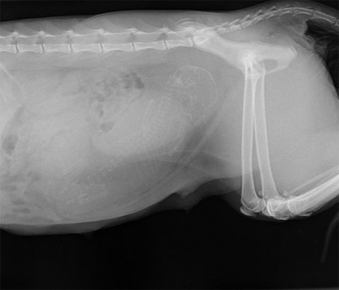
Pregnant cat with a kitten
If you look carefully in the abdomen, you can see the skeleton of an unborn kitten.

Cat who swallowed multiple hair elastics You can see both the metal joiners and the fabric of the elastic bands with the resolution of digital. Here’s the cat with his collection of hair bands.

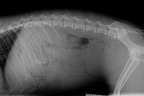
Dog who ate enough grass to fill her stomach You can see the stomach is packed full of some sort of material, food or vegetation. On endoscopy we could see it was packed with blades and stems from a coarse grass. Twelve hours of patient endoscopy and this is what we recovered, without surgery. Grass is never a square meal for your dog!